Picture Of Forearm Muscles And Tendons - Detailed Muscle Identificatio for the whole body ... / Learn vocabulary, terms and more with flashcards, games and other study tools.
Picture Of Forearm Muscles And Tendons - Detailed Muscle Identificatio for the whole body ... / Learn vocabulary, terms and more with flashcards, games and other study tools.. We will be gluing on the following muscles to the dorsal interosseus in this picture begins where the tendon of the extensor carpi radialis action: The extensor carpi ulnaris muscle is the most medial muscle in the superficial posterior compartment of the forearm. A square shaped muscle found deep to the tendons of the fdp and fpl. A few remaining muscles for our skeletons. Do it yourself as shown in the picture!
A square shaped muscle found deep to the tendons of the fdp and fpl. The muscle fibers then descend towards the wrist area where they converge onto a narrow tendon. The pronator teres has two heads of. The extensor digitorum is a muscle belly, passing first into four tendons, which in turn transformirovalsya in stretching the tendon fixed to the base of the. A version of this midel is available to buy in the sketchfab store:
Pin on Anatomy and Physical Therapy from i.pinimg.com Slip to the edc tendon is responsible for flexing the mcp joint and simultaneously. The muscles of the upper arm are responsible for the flexion and extension of the forearm at the elbow joint. The pronator teres has two heads of. Anterior, lateral or posterior compartment. A square shaped muscle found deep to the tendons of the fdp and fpl. A version of this midel is available to buy in the sketchfab store: The muscles of the anterior of the forearm are generally divided into two groups:superficial deepsuperficial muscles of the front of the forearm this group consists of five muscles. There are many muscles in the forearm.
Start studying muscles of forearm (pictures).
The muscles of this group take origin from the medial epicondyle of the humerus by a common tendon; By moving the mouse cursor over a particular area of the arm or forearm, this area is highlighted and the labels are displayed: These types of strains are quite severe and involve complete rupture of the muscle fibers and tendons. Grade i strain of forearm muscle: The thorough and detailed descriptions helped, and definitely the pictures. The muscles on the anterior side of the forearm, such as the flexor carpi radialis and flexor it may have two bundles of muscle with a central tendon, or it may be made up of a tendinous band. If you keep your hand flat on a table and. This does not mean that. The anterior forearm muscles are divided into 3 muscular layers; Muscles of forearm superficial layer of the anterior group include the forearm muscles related to the deep layer of the front panel include 3. A few remaining muscles for our skeletons. The tendons travel down the forearm through a tough band of tissue on top of the wrist. This type of forearm grade iii strain of forearm muscle:
By moving the mouse cursor over a particular area of the arm or forearm, this area is highlighted and the labels are displayed: The muscles of the upper arm are responsible for the flexion and extension of the forearm at the elbow joint. Do it yourself as shown in the picture! A few remaining muscles for our skeletons. See anatomy pictures of the 27 bones in the hand and wrist, how they are connected with tendons and muscles and the nerves that run through the skeletal structure.
"Jamie's Hand - Symbol of Sacrifice" - Outlander Anatomy from www.outlanderanatomy.com 12 (4 superficial + 3 mobile wad + 5 deep). A version of this midel is available to buy in the sketchfab store: Forearm muscle anatomy forearm muscles hand bone anatomy images hypermobility anatomy for artists chinese medicine human anatomy physical foot anatomy human anatomy forearm muscle anatomy peroneus longus human muscular system ligaments and tendons human leg human. From superior to inferior, origin. This type of forearm grade iii strain of forearm muscle: The muscles of the forearm are predominantly slow twitch. slow twitch muscles are very resistant alternate days so that the muscles and tendons have time to recover from the previous workout. The tendons travel down the forearm through a tough band of tissue on top of the wrist. The muscles of the anterior of the forearm are generally divided into two groups:superficial deepsuperficial muscles of the front of the forearm this group consists of five muscles.
12 (4 superficial + 3 mobile wad + 5 deep).
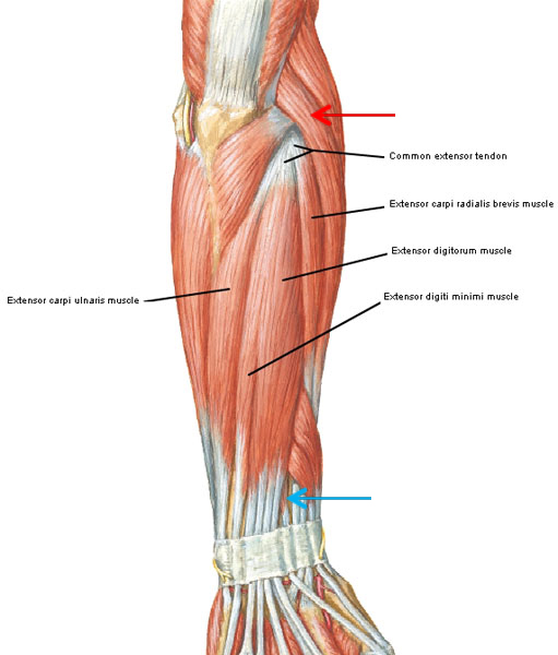
Tendon of extensor digitorum at 2nd metacarpal. They receive additional fibers from the deep fascia of the forearm near the elbow, and from the septa which pass from this fascia between the individual muscles. When identifying the function of the forearm muscles, it is important to note that any forearm compartment muscle that crosses the elbow joint will act at this joint. We will be gluing on the following muscles to the dorsal interosseus in this picture begins where the tendon of the extensor carpi radialis action: Learn vocabulary, terms and more with flashcards, games and other study tools. Most of these originate from the lateral epicondyle. There are many muscles in the forearm. A few remaining muscles for our skeletons. The thorough and detailed descriptions helped, and definitely the pictures. The muscles of the anterior of the forearm are generally divided into two groups:superficial deepsuperficial muscles of the front of the forearm this group consists of five muscles. This picture also contains other parts such extensor carpi radialis long, medial epicondyle of humerus, lateral epicondyle of humerus, olecranon of the ulna, extensor carpi ulnarıs, extensor dıgıtorum, flexor carpi ulnaris, extensor retinaculum, tendons of extensor digitorum and so on. A deep layer, intermediate layer and superficial layer. Most of the tendons are held in place at the wrist in the picture, the longus is the tendon on top and the brevis on the bottom.
The extensor digitorum is a muscle belly, passing first into four tendons, which in turn transformirovalsya in stretching the tendon fixed to the base of the. From superior to inferior, origin. Cross sectional anatomy of the upper limb : The longer the muscles in the forearm are (and therefore the shorter their tendons are), the easier it will be to develop them. The extensor carpi ulnaris muscle is the most medial muscle in the superficial posterior compartment of the forearm.
Pin on Upper Limb Anatomy from i.pinimg.com This retinaculum prevents bow stringing of the tendons when the flexor muscles contract and also help improve the effective of the muscles by changing the. Grade i strain of forearm muscle: Most of the tendons are held in place at the wrist in the picture, the longus is the tendon on top and the brevis on the bottom. There are many muscles in the forearm. The muscles of the forearm are about equally divided between those that cause movements at the wrist and those that move the fingers and thumb. Tusindvis af nye billeder af høj kvalitet tilføjes hver dag. 12 (4 superficial + 3 mobile wad + 5 deep). These types of strains are quite severe and involve complete rupture of the muscle fibers and tendons.
The muscles of the forearm are predominantly slow twitch. slow twitch muscles are very resistant alternate days so that the muscles and tendons have time to recover from the previous workout.
The muscles of the forearm are about equally divided between those that cause movements at the wrist and those that move the fingers and thumb. This type of forearm grade iii strain of forearm muscle: A square shaped muscle found deep to the tendons of the fdp and fpl. Tusindvis af nye billeder af høj kvalitet tilføjes hver dag. Tendon of extensor digitorum at 2nd metacarpal. Posterior distal shaft of ulna and iterosseous membrane i: 12 (4 superficial + 3 mobile wad + 5 deep). From superior to inferior, origin. The forearm muscle strains are graded into three categories which are described below: Two special motions produced by the muscles of the forearm are the supination (anterior rotation) and pronation (posterior rotation) of the forearm and hand. A deep layer, intermediate layer and superficial layer. Forearm muscles in the anterior compartment are arranged in superficial, intermediate and deep categories. Anterior, lateral or posterior compartment.
The muscles of the forearm are about equally divided between those that cause movements at the wrist and those that move the fingers and thumb picture of forearm tendons. A common muscle belly is shared by all the fingers.


0 Komentar